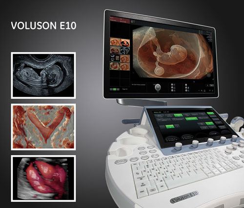
Appropriate computing capacity in its possession wonderful vistas open up before us; in addition to the wide range of diagnostic possibilities, it is very important that – since it involves post-processing of the images – the patient is not burdened with a long examination time, even if he is dressed and sitting next to us, he can review the images together with the doctor.
Some of these are:
- The images stored in the memory can be rotated in the three axes of space, so what was initially at the back, thus falling out of our field of vision, becomes clearly visible to the naked eye.
- Disturbing parts can be cut out of the images (e.g. the uterine wall can be removed from the fetus).
- Since it is a three-dimensional image, individual shadows can emphasize or obscure important details; you can adjust the shadows, but you can also add lighting to the image afterwards.
- We can choose to study the surface structures, or put them in the background and direct our attention to the bones like an X-ray.
- We can make the surface parts completely "transparent", so you can also view them in space, e.g. the brain ventricles of the fetus.
- Arbitrary surfaces that are curved in reality and therefore difficult to study can be spread out, e.g. the entire endometrium can be seen in one image. This is invaluable for infertility investigations, e.g. it provides great help in the accurate assessment of a polypus of the endometrium; the same method gives the opportunity to view the fetal spine from all directions after a few seconds of data collection ("scanning"), or to examine the vertebrae individually, without burdening the expectant mother with a lengthy examination.
- With the help of the method, we can avoid diagnostic hysteroscopy (hysteroscopy) performed under anesthesia during hospital admission, especially if a contrast material that is clearly visible with ultrasound is introduced into the uterine cavity.
Tomographic representation: during the examination, it is possible for the machine to make sections of the object to be examined with an arbitrarily defined thickness;
Flow tests: compared to traditional flow tests (Doppler), it is possible to visualize the vascular network in space; it is much easier to recognize the placenta or a possible umbilical cord knot. The vascular network of tumors can also be mapped very precisely, thus significantly facilitating the planning of surgeries.